■ 1. Background
DCLK1-IN-1 (T8418), CAS: 2222635-15-4 is a selective, in vivo–compatible chemical probe targeting the kinase domain of doublecortin-like kinase 1 (DCLK1) with high potency and low toxicity, enabling precise interrogation of DCLK1-dependent biology in cells and animal models. Quantitatively, DCLK1-IN-1 displays sub- to low-nanomolar potency by orthogonal assay formats: IC50 values of 9.5 nM (binding) and 57.2 nM (kinase assay) for DCLK1, and 31 nM (binding) and 103 nM (kinase assay) for the closely related paralog DCLK2. By engaging the catalytic domain, DCLK1-IN-1 suppresses DCLK1/2-mediated substrate phosphorylation, providing an acute and reversible means to attenuate signaling events that are otherwise difficult to isolate genetically.
DCLK1 is a microtubule-associated serine/threonine kinase harboring doublecortin (DCX) microtubule-binding motifs and a C-terminal kinase domain. It operates at the nexus of cytoskeletal organization and intracellular signaling, coordinating microtubule dynamics, cell polarity, and motility, and integrating these processes with context-dependent transcriptional programs that govern proliferation and survival. Foundational work has shown that doublecortin-like kinases regulate microtubule organization and neuronal migration, establishing DCLK-family kinases as key effectors of cytoskeletal signaling [1]. Beyond the nervous system, DCLK1 marks epithelial progenitor-like cell states linked to injury responses and neoplastic initiation, highlighting its relevance to tumor biology and regeneration [2].
Within this signaling landscape, DCLK1-IN-1 functions as a selective catalytic brake on DCLK1/2. By occupying the kinase domain and preventing phosphorylation of downstream substrates, DCLK1-IN-1 modulates pathway outputs that depend on DCLK-driven cytoskeletal control—such as microtubule stability, cargo transport, and motile behavior—and the downstream gene-expression changes coupled to these physical cues. Experimentally, this manifests as reduced levels of DCLK-dependent phosphoproteins and time-resolved alterations in microtubule-associated phenotypes. The paired potency data from binding and enzymatic assays enable calibrated target engagement across assay systems, facilitating dose selection that preserves on-target selectivity.
As a research tool, DCLK1-IN-1 enables: (i) target validation and mechanistic dissection of DCLK1/2 by acute pharmacologic perturbation, complementing genetic knockdown/knockout; (ii) phosphoproteomic mapping to define direct and indirect nodes controlled by DCLK1; (iii) deconvolution of DCLK1 versus DCLK2 contributions using differential sensitivity; and (iv) in vivo studies that link target engagement to phenotypic endpoints in tissues where DCLK1 underpins progenitor-like states and oncogenic processes [1,2]. The low-toxicity, in vivo-compatible profile supports longitudinal studies, including temporal sequencing of pathway responses and combination perturbations with cytoskeletal or growth factor pathway modulators. Collectively, DCLK1-IN-1 provides a robust chemical probe to chart DCLK-centric signaling circuits and to delineate the role of DCLK1 in cancer biology with pharmacological precision.
■ 2. Literature review
■ 2.1 Discovery of a selective inhibitor of doublecortin like kinase 1
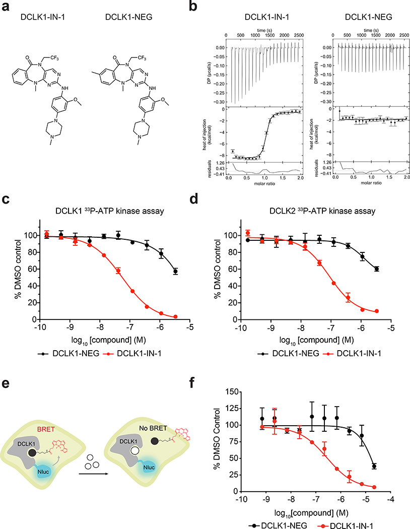
▲ DCLK1-IN-1 is a potent DCLK1/2 inhibitor. (a) Chemical structure of the DCLK1 inhibitor DCLK1-IN-1 and negative control DCLK1-NEG. (b) ITC thermograms showing the titration of DCLK1-IN-1 and DCLK1-NEG into purified, recombinant DCLK1 kinase domain. Data are representative of n = 2 experiments per compound. Integrals are shown with error bars depicting error estimates of peak integration, calculated according to ref. (c-d) DMSO-normalized results of a ³³P-labeled ATP DCLK1 kinase assay (c) and DCLK2 kinase assay (d). Assays were performed at an ATP concentration of 50 μM (DCLK1) or 100 μM (DCLK2) (Km). Data in c-d are presented as mean ± S.D. of n = 3 biologically independent samples. (e) Schematic of the DCLK1 NanoBRET assay. (f) NanoBRET evaluation of HCT116 cells expressing DCLK1-Nluc upon treatment with the indicated compounds. Data are presented as mean ± S.D. of n = 4 biologically independent samples and are representative of n = 3 independent experiments.
DCLK1-IN-1 (T8418) is presented as a selective, in vivo-compatible chemical probe of the DCLK1 kinase domain, developed through chemoproteomic profiling and structure-based design. It is described as a potent DCLK1/2 inhibitor and is shown to engage DCLK1 potently and selectively in cells. The study demonstrates activity of DCLK1-IN-1 against clinically relevant patient-derived pancreatic ductal adenocarcinoma organoid models. To characterize the cellular consequences associated with compound exposure, the authors generated RNA-sequencing datasets comparing DMSO and 2.5 µM DCLK1-IN-1 treatments for 24 hours in PANFR0172_T2, PANFR0172_T3, and PATU-8988T models. Complementary mass spectrometry-based proteomics and phosphoproteomics datasets were also produced under the same DMSO versus 2.5 µM DCLK1-IN-1, 24-hour comparison in these models. Collectively, these data establish DCLK1-IN-1 as a chemical tool and indicate its cellular target engagement and activity across cell-based and organoid contexts. The authors further state that DCLK1-IN-1 will serve as a versatile tool to investigate DCLK1 biology and to help establish the role of this kinase in cancer.[3]
■ 2.2 Inhibition of DCLK1 with DCLK1-IN-1 Suppresses Renal Cell Carcinoma Invasion and Stemness and Promotes Cytotoxic T-Cell-Mediated Anti-Tumor Immunity
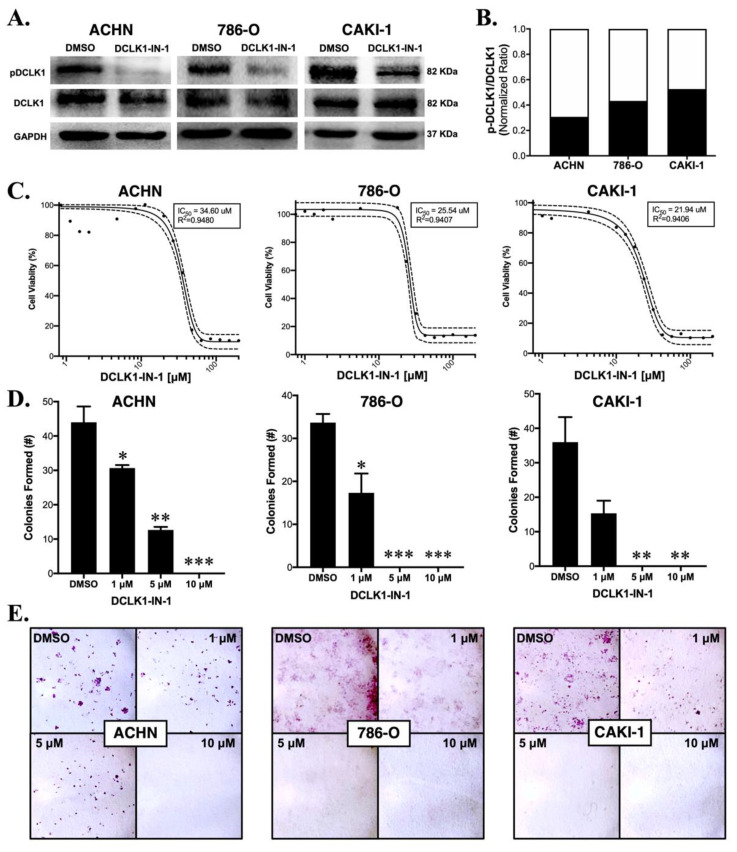
▲ DCLK1-IN-1 inhibits DCLK1 phosphorylation and colony formation in RCC cell lines. (A) Immunoblots demonstrating a strong decrease in phosphorylation of DCLK1 serine 337 by DCLK1-IN-1 (10 µM) when added to serum-starved RCC cells together with FBS for their stimulation, with incubation at the indicated time periods: ACHN (48 h), 786-O (24 h), and CAKI-1 (48 h). (B) Western blot band densitometry of phospho-DCLK1 normalized to total DCLK1 after DCLK1-IN-1 treatment (black bar) relative to dimethylsulfoxide (DMSO) vehicle control (white bar) in the ACHN, 786-O, and CAKI-1 RCC cell lines. (C) MTT cell viability assay results and IC50 curves for ACHN (48 h), 786-O (48 h), and CAKI-1 (72 h) RCC cells, demonstrating a lack of notable cytotoxic/anti-proliferative effects at concentrations ranging up to 10 µM. (D) Quantification of the mean number of colonies formed after 10 days following a single dose of DCLK1-IN-1 (1, 5, or 10 µM) or DMSO vehicle control, demonstrating a significant reduction in clonogenic capacity after DCLK1-IN-1 treatment in ACHN, 786-O, and CAKI-1 RCC cells (* p < 0.05, ** p < 0.01, *** p < 0.001 vs. DMSO). (E) Representative figures of colony formation assays quantified in (D).
DCLK1-IN-1 (T8418) is described as a small-molecule kinase inhibitor that strongly inhibits DCLK1 phosphorylation and downregulates pluripotency factors and CSC/EMT-associated markers, including c-MET, c-MYC, and N-Cadherin, in renal cell carcinoma cell lines. Functionally, DCLK1-IN-1 produced significantly reduced colony formation, migration, and invasion, and it strongly inhibited clonogenic capacity in 2D colony formation assays at doses as low as 1 μM. Consistent with effects on cancer stem-like properties, assays using floating or Matrigel spheroid protocols demonstrated potent inhibition of stemness. On the immune axis, DCLK1-IN-1 significantly reduced the expression of the immune checkpoint ligand PD-L1 in RCC cells, as shown by Western blot. In co-culture systems, DCLK1-IN-1 significantly increased immune-mediated cytotoxicity by peripheral blood monocytes and by T-cell expanded PBMCs, with dose-defined activity observed at 10 μM for naïve PBMCs and at 5 and 10 μM for T-cell expanded PBMCs; this enhancement was also evident when combined with anti-PD1 therapy. Collectively, these findings provide the first direct evidence for DCLK1-IN-1 as an immuno-oncology agent.[4]
■ 2.3 Doublecortin-like kinase 1 promotes fibroblast activation and fibrotic progression through Smad3 binding in idiopathic pulmonary fibrosis.
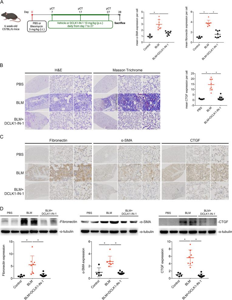
▲ DCLK1-IN-1 (10 mg/kg, oral gavage daily from day 7 after BLM administration until endpoint) attenuates bleomycin‑induced pulmonary fibrosis in mice. Histological (H&E and Masson trichrome) and immunohistochemical/Western blot analyses show that DCLK1‑IN‑1 significantly reduces fibronectin, α‑SMA, and CTGF expression compared with BLM‑treated controls (*p* < 0.05). Data are presented as means ± SEM (n=5–8 independent animals per group); one‑way ANOVA with Dunnett’s test.
DCLK1-IN-1 (T8418) is presented in this study as a selective DCLK1 inhibitor used in mice to evaluate the impact of pharmacological DCLK1 inhibition on fibrotic progression and lung function. In a bleomycin-induced pulmonary fibrosis setting, the compound was administered orally to fibrotic mouse lungs. Following administration, DCLK1-IN-1 slowed fibrosis progression and preserved lung function, demonstrating a beneficial effect on disease course within this experimental model. The study explicitly positioned DCLK1-IN-1 to probe how targeted kinase inhibition influences fibrotic remodeling and respiratory performance, and the outcomes indicated that pharmacological inhibition through DCLK1-IN-1 produced measurable improvements in the assessed functional and fibrotic endpoints. Across the in vivo experiments, the positive readouts tied directly to oral delivery of DCLK1-IN-1 in bleomycin-treated mice, and the reported effects encompassed both attenuation of fibrotic progression and maintenance of lung function metrics. Collectively, these findings describe DCLK1-IN-1 as an orally administered, selective inhibitor that, within the bleomycin fibrosis model, exerted protective effects on the progression of fibrosis while preserving functional respiratory capacity. The observations were derived from comparisons within the bleomycin-challenged cohorts, supporting a role for DCLK1-IN-1 in moderating fibrosis-related pathology and sustaining lung function in this preclinical context.[5]
■ 2.4 DCLK1 isoform (DCLK1-S) as a critical player in promoting inflammation, tissue remodeling, and EMT in mouse models of colitis.

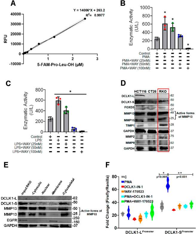
▲ WAY 170523, a selective MMP13 inhibitor, dose‑dependently (25–100 nM) reduces MMP13 enzymatic activity in PMA‑ or LPS‑stimulated RKO cells (B, C). Among colon cancer cell lines, RKO shows higher protein levels of DCLK1‑L, DCLK1‑S, and MMP13 (D). Subcellular fractionation reveals distinct distribution of MMP13 (mainly cytosolic and membrane) and DCLK1 isoforms (E). Dual‑luciferase promoter‑reporter assays demonstrate differential promoter activities between DCLK1‑L and DCLK1‑S (F). Data are mean ± SEM, n=3 independent experiments; *p<0.05, **p<0.005, ***p<0.001.
DCLK1-IN-1 (T8418) is identified as a DCLK1 inhibitor and was evaluated in promoter-reporter assays focused on DCLK1 isoforms. In HEK293 cells transfected with DCLK1 promoter constructs, cells were exposed to PMA to stimulate promoter activity and then treated with DCLK1-IN-1 either alone or in combination with PMA. Under these conditions, DCLK1-IN-1 decreased activity driven by the DCLK1-L promoter. When the DCLK1-S promoter was assessed, PMA-induced increases in promoter activity were irreversibly inhibited by DCLK1-IN-1 whether applied by itself or together with PMA. Across assays comparing the two isoform promoters, DCLK1-IN-1 produced a more dramatic decrease in DCLK1-S reporter activity than in DCLK1-L, highlighting a comparatively stronger impact on the short isoform’s transcriptional reporter signal. Collectively, within this experimental system, DCLK1-IN-1 functioned as an inhibitor that suppressed reporter activity from DCLK1 promoters, with the most pronounced effect observed on the PMA-stimulated DCLK1-S promoter.[6]
■ 3. Conclusion
DCLK1-IN-1 functions by selectively binding to the catalytic kinase domain of DCLK1 and DCLK2, thereby blocking substrate phosphorylation and acutely inhibiting DCLK-dependent signaling pathways involved in cytoskeletal control and gene expression. Literature reports demonstrate its potent and selective inhibition of DCLK1/2 in cellular and animal models, effectiveness in suppressing cancer stemness, invasion, and enhancing immune-mediated tumor clearance, as well as its ability to slow fibrotic progression and preserve lung function in pulmonary fibrosis models. Additionally, DCLK1-IN-1 modulates promoter activity of DCLK1 isoforms linked to inflammation and tissue remodeling. Future applications include detailed mechanistic studies of DCLK1 function, target validation in oncology and fibrotic diseases, phosphoproteomic mapping of signaling networks, and exploration of combination therapies with immune checkpoint inhibitors and cytoskeletal modulators.
■ 4. Q&A
Q1: What is the mechanism of action of DCLK1-IN-1?
A1: DCLK1-IN-1 selectively binds the kinase domain of DCLK1 and DCLK2, blocking substrate phosphorylation and thereby acutely and reversibly inhibiting DCLK-dependent signaling pathways related to cytoskeletal dynamics and gene expression.
Q2: What key biological effects of DCLK1-IN-1 have been demonstrated in cancer models?
A2: DCLK1-IN-1 suppresses pluripotency factors and CSC/EMT markers, reduces colony formation, migration, and invasion, inhibits stemness in spheroid assays, downregulates PD-L1 expression, and enhances immune-mediated cytotoxicity, including synergy with anti-PD1 therapy.
Q3: How has DCLK1-IN-1 been used in fibrosis research?
A3: In a bleomycin-induced pulmonary fibrosis mouse model, oral administration of DCLK1-IN-1 slowed fibrosis progression and preserved lung function, demonstrating beneficial effects on fibrotic remodeling and respiratory performance.
■ 5. Reference
[1] Koizumi H, Tanaka T, et al. Doublecortin-like kinase functions with doublecortin to regulate microtubule organization and neuronal migration. Neuron. 2006;49(1):55-66.
[2] Westphalen CB, Takemoto Y, et al. Dclk1 Defines Quiescent Pancreatic Progenitors that Promote Injury-Induced Regeneration and Tumorigenesis. Cell. 2016;166(2):358-371.
[3] Ferguson F, Nabet B, Raghavan S, Liu Y, Leggett A, Kuljanin M, et al.. Discovery of a selective inhibitor of doublecortin like kinase 1. Nature Chemical Biology. 2020;16(6):635-643.
[4] Ding L, Yang Y, Ge Y, Lu Q, Yan Z, Chen X, et al.. Inhibition of DCLK1 with DCLK1-IN-1 Suppresses Renal Cell Carcinoma Invasion and Stemness and Promotes Cytotoxic T-Cell-Mediated Anti-Tumor Immunity. Cancers. 2021;13(22):5729.
[5] Lin L, Cheng W, Wu Y, Wen H, Hsu H, Liu C, et al.. Doublecortin-like kinase 1 promotes fibroblast activation and fibrotic progression through Smad3 binding in idiopathic pulmonary fibrosis.. Journal of biomedical science. 2026;33(1):.
[6] Yusuf K, Roy B, Hauser W, Al-Kasspooles M, Sanchez S, Attard T, et al.. DCLK1 isoform (DCLK1-S) as a critical player in promoting inflammation, tissue remodeling, and EMT in mouse models of colitis.. PLoS pathogens. 2025;21(8):e1013360.
https://www.targetmol.com/
TargetMol Chemicals Inc.
About Author